

Suspensory Ligament Injuries: Advances in Diagnosis and Treatment

Updated:
November 6, 2013By Mike Scott, DVM, MVSc
Suspensory ligament injuries are a common cause of lameness in the horse, particularly athletic horses and those involved in competitive events. Often these injuries are chronic and have a high probability of reccurrence, which makes them a significant concern for horse owners.
Recent advances in our understanding of these injuries, improvements in diagnosis, and new options for treatment have improved the prognosis for horses with these injuries. In this article, we’ll review the anatomy and physiology of the suspensory ligament and present current concepts on diagnosis and treatment of suspensory ligament injuries.
Anatomy & Physiology
The anatomy of the lower limb of the horse below the knee or hock is very similar. The horse has evolved to the point where it is walking on what would be the equivalent of our middle or third finger, with the joints of the knee or hock being the equivalent of our wrist (carpus), or ankle (tarsus). The weight of the horse is borne on a central column of bones comprised of the cannon bone (third metacarpus or metatarsus), the pastern bones, and the coffin bone within the hoof. This column of bones is supported by several large tendons and ligaments in the back of the leg including the superficial digital flexor tendon, the deep digital flexor tendon, and the suspensory ligament.
Horses have evolved and adapted to move rapidly and efficiently. Using principles of kinematics one can calculate theoretical limits for the speed a horse should attain when galloping based on the mass of its muscles, the dimensions of its skeleton, and the amount of force it should be able to produce to propel its body mass forward.
Interestingly, when we compare actual measurements of athletic performance to calculated estimates the horse consistently exceeds the estimates by 10 to 20 percent.

Anatomy of the Lower Limb (Front Leg)
Part of the reason for this is the fact that the flexor tendons and suspensory ligament act like giant rubber bands to store and return kinetic energy to the galloping horse. Muscle contraction can only change the length of the superficial flexor tendon by two percent, whereas it has the ability to repeatedly stretch under load an additional 16 percent, and return to normal length without injury.
The function of the suspensory ligament is more complex than simply acting as a passive connector between two bones, as the traditional concept of a ligament describes. Its principal role is to prevent excessive extension of the fetlock joint. It also acts in the capture and release of kinetic energy and in the dynamic support of the limb during athletic performance.
It is useful to think of the suspensory ligament in three separate regions: the upper (proximal) part, the body, and the branches. It is approximately 10 to 12 centimeters long and approximately one square centimeter in cross-sectional area. It originates at the proximal aspect of the cannon bone and inserts on the sesamoid bones located at the back of the fetlock joint. The body of the suspensory ligament descends between the two splint bones, which shield the upper half of the ligament on either side. The ligament is essentially shaped like an upside-down Y, with a division somewhere at the mid-cannon level and one arm of the Y attaching to each sesamoid bone. Below the level of the fetlock joint, the limb support functions of the suspensory ligament are continued by the sesamoidean ligaments.
Injury
An important concept to realize is that suspensory ligaments seem to be affected by different types of injuries. Injury to the upper third of the suspensory ligament and the origin is usually called proximal suspensory desmitis (PSD). PSD of the forelimbs is different from that affecting the hind limbs, with significant differences in prognosis and approaches to treatment. Injury to the body of the suspensory ligament is principally an injury of racehorses and jumping horses. Injury of the suspensory branches is relatively common in all types of sport horses.
Suspensory ligament injuries may be singular traumatic events associated with an accident or hyperextension, they may be repetitive strain injuries, or they may be associated with chronic degeneration of the ligament over time.
We do not really have a good understanding of the mechanisms of injury of this tissue, but it does seem to be more complex than simply “stepping in a gopher hole.” As our understanding of genetics improves for both human and animal patients, there is increasing evidence that variation within individuals exists which predisposes to injury. Trauma is often involved, but predisposing intrinsic factors may also influence risk.
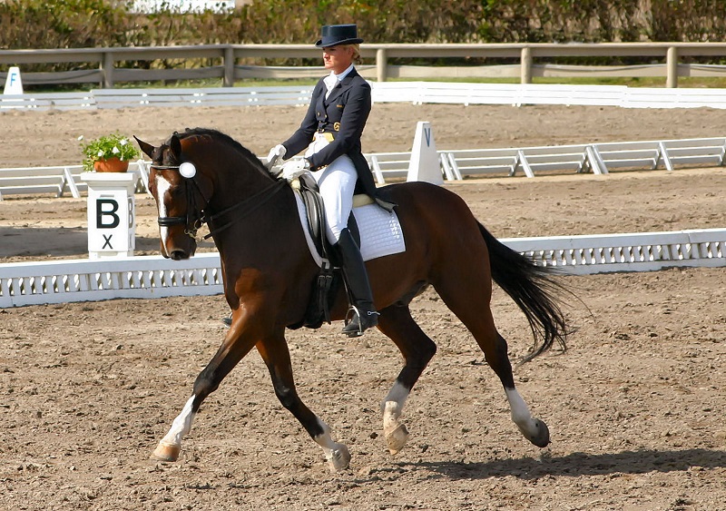
Horses performing extreme athletic efforts such as jumping and racing are at increased risk for suspensory ligament injuries, as are horses that perform repetitive movements, such as upper-level dressage horses. Photo: Shutterstock/Michelle Marsan
Environmental factors that contribute to suspensory injuries include the nature of work, the shoeing of the horse, and the footing on which the horse is worked. Horses performing extreme athletic efforts such as jumping and racing are at increased risk. Horses that perform repetitive movements also seem to be at risk, such as more advanced level dressage horses and cutting horses. There may also be a combined risk for young (two and three-year-old) horses performing repetitive movements. Foot balance likely plays a role in predisposing horses to suspensory injury. A steep foot angle increases the strain on the suspensory ligament, and medial to lateral imbalance may predispose to suspensory branch injuries. It seems that deep footing or slippery footing also increases the risk of these types of injuries.
Diagnosis
Although injuries to the suspensory ligament are relatively common, often they are not diagnosed, or are misdiagnosed for a period of time. There are several reasons for this. Frequently there is little or no visible swelling in the limb, so there are no physical clues. The lameness may be mild and sporadic, often resolving after a short period of rest and returning after periods of work. Horses are often affected bilaterally, which means that they gain little advantage by limping and instead just shorten their stride and appear weak or don’t perform well. Often when lameness examinations are performed, little to no lameness is seen and nothing definite is seen on radiographs. Finally, horses with suspensory injuries may show a transient response to joint injections or systemic medications, which may lead us to believe that the problem is joint related. A common presentation for bilateral PSD in the hind limbs would be a horse that has decreased performance, does not really appear lame, has a sore back and sacro-iliac region, and has shown transient (approximately two weeks) improvement after hock joint injections.
A proper lameness examination is the first step in diagnosing suspensory ligament injuries. In some cases, such as suspensory branch injuries, there may be swelling or pain on palpation present, but in many cases the physical symptoms are mild or absent. Dynamic examination should be performed by watching the horse walk and trot on a straight line and walk, trot, and canter circles in each direction.
Often it helps to examine the horse on both hard and soft footing as it seems that they may actually look worse on softer footing, and with the affected limb on the outside of a circle. In some cases watching the horse under saddle is helpful as the lameness may be more visible with a rider up.
It is usually essential to perform diagnostic nerve blocks to determine the source of pain, particularly in the diagnosis of PSD. Diagnostic imaging cannot show where the pain comes from; it is important to confirm that when the region of the suspensory is frozen, the horse’s gait changes (improves). This is usually the most important step in the diagnosis of these injuries. Once there is evidence that the region of the suspensory ligament is the source of pain, the logical next step is to perform diagnostic imaging of this region.

A thorough lameness examination includes walking and trotting your horse in hand on a straight line for your veterinarian to observe. Photo: Robin Duncan Photography
Ultrasonography is usually essential in the diagnosis of suspensory ligament injuries. Abnormalities seen on ultrasound include enlargement of the ligament, bone abnormalities at the origin and insertion, and abnormalities in the appearance of the fibre pattern within the ligament or possible evidence of fibre disruption, typically seen as darker areas within the body of the ligament.
In a recent study comparing ultrasound and magnetic resonance imaging (MRI) examination of horses with hind limb PSD, ultrasonography identified the injury in approximately 75 percent of cases compared to 100 percent for MRI.
MRI examination provides the best method of imaging the suspensory ligament and the best means of making an accurate diagnosis and determining the exact nature and severity of injury. The availability of MRI for horses is gradually increasing, and our understanding of what we see on these images is becoming more meaningful.
Treatment & Rehabilitation
Rest, Ice, Compression
Rest is the foundation of treatment of suspensory ligament injuries, regardless of their location. The pain associated with suspensory ligament injuries is often transient and short lived. A short time after injury, the horse may look and feel “better” and may be returned to work only to have the lameness return.
A rest period of three months would be typical for relatively moderate injuries. Horses with more chronic or severe injuries may require longer periods of time off, in some cases approximately a year. Rest does not mean turnout in a field; rest means confinement to a box stall or a small paddock with controlled exercise such as hand walking.
Horses with PSD of the forelimbs have a fairly good prognosis, with many horses responding to rest within three or four months. In contrast to this, horses with PSD of the hind limbs have a poor prognosis with rest alone. In one study, only 14 percent of horses with PSD of the hind limbs were able to return to work and remain sound. Injuries of the suspensory branches generally respond fairly well to rest alone, although some horses will require nine months or more before being able to return to work safely.
In the acute stages (the first 10 to 14 days) of any type of suspensory ligament injury, it makes sense to apply cold and compression to the injured region in an attempt to minimize swelling and pain.
Peri-ligamentous Injections
In cases of acute injury where there are no definite ultrasonographic abnormalities of the ligament, the horse may respond reasonably well to local infiltration of anti-inflammatory medications such as corticosteroids or autogenous orthokine serum (IRAP). The purpose of this treatment is to minimize inflammation and swelling of the ligament, which may alleviate pain and further damage. In the proximal portion of the limb, the suspensory ligament is bounded on three sides by bone (the cannon bone and the medial and lateral splint bones) and on the fourth side by a tough sheet of dense tissue called fascia. It is essentially contained within an unyielding “box.” In theory, if the ligament sustains injury and begins to swell, the pressure in the “box” increases, creating what is called a compartment syndrome. Compartment syndromes are typically painful, and may lead to tissue damage and delayed healing due to the pressure in the compartment limiting blood flow to the area. Reducing inflammation may prevent or reduce the severity of potential compartment syndrome.
Shock Wave Therapy
Shock wave therapy (SWT) appears to be helpful in the treatment of suspensory ligament injuries, both in reducing the time needed before returning to work and reducing the rate of re-injury. In one study, approximately 50 percent of horses with chronic PSD in the front or hind limbs returned to work after six months rest and treatment with SWT. Although SWT appears to affect tissue healing responses, the mechanism(s) of action remain unclear.
Ligamentous Injections
When an area of injury is visible on ultrasonographic examination, the question frequently arises regarding the benefit of injecting a substance into the area in an attempt to affect the healing response. Over the past several decades a variety of substances have been used for this purpose, often with mixed results or no results. Currently the focus is on using biologically based products such as bone marrow, plasma products, or cell cultures in an attempt to create a condition of tissue regeneration rather than just tissue repair.
Bone marrow collected from the patient has been used to inject into areas of injury with the goal of introducing cells and growth factors which may be regenerative or at least improve the quality of repair. This treatment was reported to have some success, but also may be associated with complications such as calcification of the ligament.
Autologous (meaning “from the self”) stem cell injections may be a beneficial treatment option for suspensory ligament injuries. After injury, the scar tissue that replaces the damaged ligament may result in reduced performance and increased risk of re-injury. If regeneration of the injured tissue can be achieved rather than scar tissue-based repair, it may be possible to restore more of the normal structure architecture and biomechanical function of the tissue. This is the basic premise behind the concept of “regenerative” medical treatments including stem cell treatments.

Rest, consisting of confinement either to a box stall or a small paddock, is essential to the treatment of suspensory ligament injuries.
At this point much of this is theoretical, and controlled trials proving these theories are still lacking.
Platelet rich plasma (PRP) is a serum fraction made from the horse’s own blood, which contains a concentrated volume of platelets. Platelets are tiny cell fragments (cells that do not contain a nucleus) normally found in the blood. They have critical functions in blood clotting, and are a natural source of growth factors (chemical mediators of tissue growth, repair, and regeneration). PRP can be manufactured from the horse’s own blood and used to treat suspensory ligament injuries, alone or in combination with other therapies. Although controlled studies are lacking, in one report on the use of PRP to treat mid-body suspensory ligament injuries in racehorses, all horses returned to racing and had excellent results.
Surgery
Horses affected with proximal suspensory ligament injuries in the hind limb appear to have the worst prognosis, and often suffer from chronic lameness or re-injury after returning to work. The prognosis remains poor even with other forms of treatment. This challenge has led to the development of surgical treatment options:
- Desmoplasty and Fasciotomy: This treatment involves making stab incisions into the upper portion of the ligament, making a small incision into the tough sheet of tissue surrounding the back of the ligament (fasciotomy) and incising the tissues of the ligament itself (desmoplasty). In theory, this procedure may release pressure in the compartment surrounding the injured ligament, and may stimulate a repair response in the chronically injured ligament. In one study of 27 horses, 85 percent of those treated returned to work after surgery and rehabilitation.
- Fasciotomy and Neurectomy: This surgery involves making an incision in the hind limb to access the proximal suspensory ligament, and then cutting the deep branch of the lateral plantar nerve (neurectomy) and the tough sheet of tissue surrounding the back of the suspensory ligament (fasciotomy). The nerve branch that is cut provides sensation to the upper portion of the suspensory ligament; cutting this branch reduces pain perception in the ligament but has no effect on the other regions of the leg. In a study of 20 horses treated in this way, 19 returned to their previous level of exercise with only two horses experiencing a recurrence of PSD. In our practice I am currently combining this surgery with injection of bone marrow derived stem cells or PRP into the injured regions of the suspensory ligament.
Rehabilitation
The rehabilitation of the horse with tendon and ligament injuries is a complex process that is not well understood. At this point the main principles guiding this process are adequate periods of rest and gradual, controlled exercise of increasing duration and intensity. Humans training for athletic endeavours frequently use a guiding principle of only increasing the intensity and duration of work by 10 percent per week or less to avoid injury. Similar guidelines should be used for horses but the details are not well defined and are often based on guesswork. Using repeated ultrasound examinations to assess ligament healing is helpful, but in some cases the injured tissues always remain ultrasonographically abnormal so there are limitations to the information obtained.
There has been an increase in the availability of rehabilitation facilities with various types of equipment and techniques for rehabilitating horses, including treadmills, aqua-treadmills, swimming pools, cold water baths, and automatic walkers. There is a remarkable lack of good information on how these modalities should be used. Studies are currently being performed to assess the benefits of these rehabilitation methods and to determine their advantages and disadvantages.
Additional complementary therapies such as magnetic devices, therapeutic ultrasound, light therapy, acupuncture, chiropractic, and others all seem to provide some benefit in certain situations but again the science guiding their application is lacking. A good general principle is to rely on an accurate diagnosis and regular recheck examinations to determine the exact nature of injury initially and to guide progressive steps in the rehabilitation process.
Prognosis
The prognosis for horses affected with suspensory ligament injuries varies with the severity, chronicity, and location of the injury. Minor, acute injuries can have a good prognosis and only require a relatively short rest period, while severe, chronic injuries can cause such pain that euthanasia is the most humane option for the horse. The best treatment options and prognosis can only be determined after a careful lameness examination has been performed and an accurate diagnosis has been made. Recent and ongoing research into the nature of suspensory ligament injuries and their diagnosis and treatment should help in treating these cases more successfully in the future.
Mike Scott, DVM, MVSc, is a Diplomate of the American College of Veterinary Surgeons, and a Board Certified Specialist in Large Animal Surgery. His practice is based at Moore Equine Veterinary Centre Ltd. on the north edge of Calgary, Alberta. www.mooreequine.ca
Main Article Photo: Robin Duncan Photography
This article originally appeared in the June 2012 issue of Canadian Horse Journal.
Blogs & Podcasts

Advertisement

Related Articles


Advertisement

Featured Stories


Advertisement
