

White Line Disease

Updated:
March 13, 2014By Kentucky Equine Research
The white line is the narrow, light-coloured band visible on the underside of a freshly trimmed hoof at the junction of the hoof wall and the sole. White line disease, an infection that causes separation of the wall, may be seen first at the white line but actually affects the zone of contact between the hard outer hoof wall and the middle layer of hoof tissue. It occurs most commonly in front feet but can occur in any foot. The old saying, “No foot, no horse” might well have been coined in reference to this serious problem, which can lead to months of intensive treatment. In severe cases, lameness may develop. Left untreated, white line disease can result in rotation of the coffin bone due to widespread damage in the supportive structures of the hoof.
Causes
White line disease has been recognized for years and has accumulated names such as seedy toe, hollow foot, wall thrush, and stall rot. Despite its long history, the unknown factors exceed what is understood about the condition. It is not clear, for instance, whether the cause is fungal or bacterial, or possibly a combination of the two. Anaerobic organisms have been found in affected tissue, but aerobic strains have not been eliminated from consideration. It has been suggested that some type of stress (faulty hoof conformation, flexor deformities, concussion on hard ground, leverage created by overly long toes, or the combination of an overweight horse and small hooves) causes the laminae to tear and bleed. This trauma may provide a growth center for soil-dwelling bacteria or fungi that enter the hoof through cracks or nail holes.

Horse hoof anatomy
Visible on the underside of a freshly trimmed hoof, the white line is the narrow, light yellowish band between the hoof wall and the sole.
Other factors that have been vaguely connected with white line disease are lack of exercise, shoes that are too small, poor stable hygiene, wet pastures or stalls, and inadequate nutrition. For every hypothesis, however, there seem to be cases that do not fit the profile.
Diagnosis
White line disease may be diagnosed during a routine trimming when a farrier notices a small area of crumbly or powdery black or gray tissue at the white line. Paring away the damaged horn reveals separation of the hoof layers leading upward from the toe toward the coronary band. Tapping on the hoof wall over the separation produces a hollow sound. Bulges or sunken areas of the hoof are sometimes noticed. The horse is usually not lame at this stage.
Treatment
Owners will need to consult both a veterinarian and a farrier for help in eliminating white line disease. Treatment usually involves four steps. First, all infected tissue must be removed. This can sometimes be accomplished by reaching into the hollow area with a hoof knife and scraping until healthy tissue is encountered. In more advanced cases, the hoof wall over the affected part is removed. Problems in resolving the condition are often blamed on failure to eliminate every bit of infection. A farrier may need to examine the horse as often as every ten days to cut out areas that show damage.
The second step is application of an antibacterial or antifungal product. Iodine, bleach, hydrogen peroxide, copper sulfate, and a long list of commercial hoof disinfectants have been used with varied success. Anecdotal evidence to the contrary, no one product seems to be effective in every case. A veterinarian's recommendations can help an owner choose a product that will stop the infection without damaging healthy tissue. Surprisingly, there is evidence that some cases of white line disease resolve after thorough removal of all affected tissue without use of disinfectants.

Photo: Olds College/Dean Sinclair, CJF
Despite its name, white line disease actually affects the hoof tissue between the middle and outer layers of the hoof wall, causing separation of the wall.
The next phase involves keeping the hoof from becoming re-infected. Exposure to air and avoidance of moisture are both important factors. Although some treatments involve packing the separated area with medicine or covering the affected hoof with an acrylic patch to prevent entry of dirt and moisture, these measures have sometimes been blamed for perpetuating the dark, damp conditions that allow the infection to persist and spread.
The final step in the treatment of white line disease is protection during new horn growth, a process that takes several months to a year. Shoeing helps support the hoof and keep pressure off the toe. Depending on the location and amount of wall that has been removed, it may be necessary to use egg-bar or heart-bar shoes, glue-on shoes, or shoes with extra clips or screws. In many cases the infection is quite difficult to eliminate, and even in horses that seem to recover completely, recurrence is frequently seen within a few years.
Prevention
No horse is immune to hoof problems, but common sense implies that this infection can be minimized by maintaining clean stalls, scheduling regular farrier visits, and providing balanced nutrition. A study at the University of Edinburgh confirmed that prolonged contact with manure caused disintegration of hoof tissue, and the damage was especially severe when the hoof wall was already in poor condition. Trimming or resetting shoes every five to six weeks protects the hooves in two ways – the tissue strain caused by imbalances or long toes can usually be avoided, and frequent exams allow a farrier to find evidence of disease before the infection has destroyed extensive areas of the hoof.
The dietary element of managing white line disease involves feeding recommended amounts of energy, vitamins, and minerals. A hoof supplement that provides additional biotin, methionine, zinc, and iodine will supply nutrients that are essential for the development and maintenance of strongly bonded layers of hoof tissue.
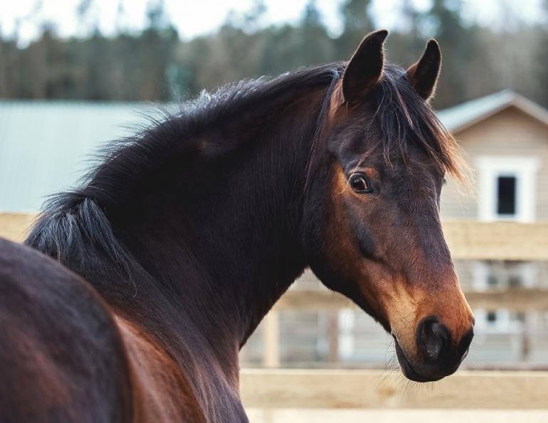
Main Article Photo: Olds College/Dean Sinclair, CJF - Treatment for white line disease begins with the paring or scraping away of the infected hoof tissue. Advanced cases may require complete removal of the hoof wall over the affected area.
This article appeared in the August 2013 issue of Canadian Horse Journal.


Blogs & Podcasts

Advertisement
Related Articles


Advertisement

Featured Stories


Advertisement
