
Your Horse's Heart
Updated:
November 14, 2014Skipped Beats, Sudden Death… and Why We Shouldn’t Worry Too Much
By Peter W. Physick-Sheard, BVSc, MSc, FRCVS
When you first start examining patients as a veterinary student, you’re very keen to (gently) poke and prod every animal you come across. Realizing you can assess cardiovascular function by palpating peripheral pulses is very empowering!
Once you find a pulse in a healthy cow, you simply hang on and count, as the pulse waves come to you in a more or less steady stream, 60 to 80 times a minute. You can confidently anticipate when the next one is going to arrive. Then you examine a horse, perhaps a mare in her late teens – quiet, cooperative, and relaxed - and all of your confidence disappears. One minute you have the pulse, the next you don’t. One minute the pulse is strong, then it disappears – and you’re sure you didn’t move your fingers. The book says the rate should be 28 to 40 beats per minute, but in this horse sometimes it’s 40 and sometimes it’s 12.
Horses have a wide range of normal resting heart rates and a huge range of different normal rhythms, and none of them are regular in a healthy horse. Photo: Just Chaos/Flickr
Generations of veterinary students have gone through this – examining supposedly healthy horses whose hearts do not beat regularly, horses whose hearts take frequent breaks, beat much more slowly, and sometimes much more rapidly, than they are supposed to. The horse has a huge range of normal resting heart rates and a huge range of different, normal rhythms, and in a healthy horse none of them are really regular. This is your introduction to equine cardiology – all over the map!
Cardiovascular Reserve and the Microcirculation
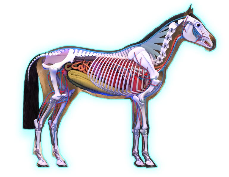
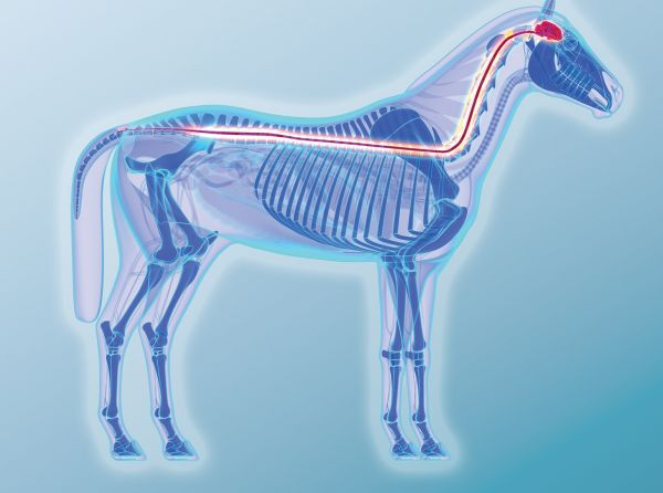
The cardiovascular system of the horse consists of a heart, the arteries that convey cardiac output (blood) to functioning tissues, the microcirculation inside those tissues that moderates local distribution of blood flow, and the venous system that collects blood from tissues and returns it to the heart. If I were to ask you which part of this system is most important, which part is in charge, you would probably say the heart – but that’s not true. It’s actually the microcirculation.

The equine heart is a muscular pump that circulates blood around the body from the area of highest pressure (the left ventricle) to the area of lowest pressure (the right ventricle).
The cardiovascular system exists for no reason other than to meet the needs of tissues, muscle for example, and in this regard, the heart is an essential but slavish bag of muscle, a pump. It goes like this: blood flows from areas of high pressure to areas of low pressure. The highest pressure is at the left ventricle of the heart during systole or contraction, where the pressure is generated, and it’s lowest in the right ventricle in diastole or relaxation, when the right ventricle is filling - blood flows around the body from the left ventricle to the right ventricle. In the middle of the circulation, between those two ends, lies the microcirculation, which supplies functioning tissues with the oxygen and fuel they need and removes waste products to transport them to where they can be disposed of.
With the exception of extreme circumstances, such as severe shock, the tissue or peripheral circulation functions entirely autonomously – without any reference whatsoever to what is going on centrally in the heart. Small arteries called arterioles lying at the beginning of the microcirculation are controlled almost exclusively by local factors such as potassium levels, oxygen tension, and acidity, and these are determined by local metabolic activity. So long as there is adequate pressure to drive flow, local arterioles will ensure tissues get what they need. The role of the heart is simply to maintain the pressure.
If maintaining pressure, and thus flow, is the primary role of the heart, how might pressure change, and how is that change monitored? Moment-to-moment control of blood pressure is determined by special pressure sensitive tissues distributed throughout the vascular system. If pressure falls, these sensors send a message to the brainstem that results in an increase in heart rate and thus output. In the horse, this is initially achieved by reducing activity in a branch of the autonomic nervous system, the parasympathetic system, which reduces inhibition of the heart. This works because in the horse at rest, the heart rate is actually being held down by the parasympathetic activity.
At the same time, adjustments in return of blood from peripheral tissues, in pressures in the central venous reservoir (the large veins leading to the heart), and in contractility or responsiveness of the heart itself are taking place. Such mechanisms conspire to promote an increase in filling of the heart (so filling will be efficient at the higher heart rate), to support the increased output.
Exercise
Progressive increase in the amount of muscle tissue functioning vigorously as exercise intensity increases will be associated with progressive dilation of all the arterioles in that muscle, and with filling of the muscle capillary bed (all of the thin-walled, microscopic vessels fed by the arteriole and that supply nutrients directly to the tissue). This shift in blood volume from the largest veins to the capillaries, and the fall in blood flow resistance that results from dilating arterioles will result in a fall in blood pressure if not properly managed. The more those peripheral resistance vessels (the arterioles) dilate, the more “leaky” the system becomes, and the harder the heart must pump to maintain pressure. As demand continues to increase with increasing work, there is a progressive withdrawal of parasympathetic nervous inhibition and a simultaneous progressive increase in sympathetic nervous system activity. The latter has numerous effects, including increasing heart rate, increasing heart muscle contractility, optimising venous return and cardiac filling, and redistributing blood flow through tone in major muscular arteries to favour flow to functioning tissues.
Normal Rhythm – How Normal is Normal?
What does this have to do with heart rhythm? First, there's no such thing as a steady heart rate. An absolutely steady, metronome-like rhythm is not normal. Normality involves constant change and readjustment - the ability of the body to respond rapidly and appropriately to these changes is in fact an index of health. If you monitor the electrocardiogram (ECG) of a horse at complete rest and measure fluctuations in the interval between consecutive beats (called instantaneous heart rate), you get what is called a heart rate time series. In a normal animal this is anything but a straight line. The amount and pattern of variation from beat to beat (called Heart Rate Variability, or HRV), changes tremendously from horse to horse, circumstance to circumstance, moment to moment.
Analysis of this sequence of instantaneous heart rates helps demonstrate that underlying control mechanisms, such as blood pressure control for example, don’t simply turn on or off when necessary. Instead, they tick along continuously in a cyclic or periodic manner at varying frequencies. When changes in blood pressure occur, the amplitude and frequency of these control cycles change, and this happens constantly. You can relate these cycles to the balance in underlying autonomic nervous system (parasympathetic and sympathetic) control.
Back to the mare being examined by the veterinary students. She is quiet and relaxed, almost sleeping. She has very little sympathetic activity and lots of parasympathetic activity. This means she will have lots of high frequency changes in instantaneous heart rate, making adjustments with every beat. Occasionally this will result in her dropping a beat completely; at other times the interval between beats will be variably long. You would imagine that if everything was completely stable, she ought to maintain the same slow basic rate, but it’s not that simple, because blood pressure is not the only thing stimulating her parasympathetic nervous system.
She is also breathing, very slowly (and deeply, maybe six to eight breaths per minute), and these breathing cycles are also adjusting both blood pressure and parasympathetic activity. As a result, the actual effect on heart rate (beat-to-beat) sometimes adds to, and sometimes subtracts from other influences. Since the input of the parasympathetic nervous system to the heart actually has a left and right branch, and since those branches tend to innervate different parts of the heart, there are also different options to slow down the heart and reduce cardiac output. These are by slowing down the primary pacemaker (the sinus node), the atrioventricular (AV) node, which lies between the atria and ventricles in the middle of the heart’s conduction system, or by using both. The result is a wide variation in instantaneous heart rate and rhythm.

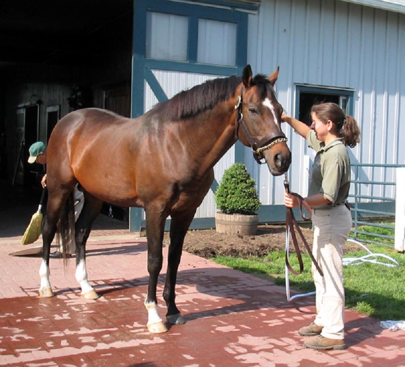
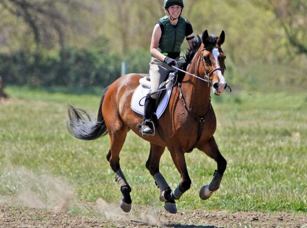
The resting heart rate of a fit horse with several years of aerobic training may drop two heartbeats at a time, and may be a slow 16 beats per minute. From prolonged training, the horse will have a larger heart which can perhaps put out enough blood in three consecutive beats to take the next two off. The result is a huge variation in resting heart rate, and it’s all normal. Photo: John Shortland/Flickr
Consider a fit event horse with several years of aerobic training. At rest, it sometimes drops two beats at a time, and its resting heart rate is 16 beats per minute. Yes, the horse is very relaxed, but it also has a large heart both by virtue of genetic selection and prolonged training. It can perhaps put out enough blood in three consecutive beats to take the next two off. The result is a huge variation in resting heart rate, and it’s all normal.
Can such low heart rates at rest ever be abnormal? Yes, but typically there would be other signs telling you if something was wrong, and such circumstances are rare. There are many other variations on “normal” encountered at rest, but few represent a problem of any significance. All mammals also throw occasional extra beats, for example, and these may come from the atria or ventricles. So long as every extra beat comes from the same place in the heart, one extra beat every couple of minutes is regarded by many clinicians as normal and of no concern.
What IS Abnormal?
There’s a small group of rhythm disturbances that come from the atria and are clearly a problem. These are third-degree heart block, atrial fibrillation, atrial flutter/tachycardia, and frequent atrial ectopic beats arising from multiple foci with random timing. None of these disturbances will disappear with stimulation and all have a performance impact; however, it’s unlikely they would lead to death, and with the exception of atrial fibrillation, they are not common. In fact, some of these may not be noticed unless the horse is examined by a vet or is asked for a maximal performance. Atrial fibrillation is quite common and can be treated in most cases. Third degree heart block is thankfully very uncommon, and usually untreatable. Atrial flutter is also very uncommon, as are complex atrial ectopic beats, but in horses they can often be managed.
Ventricular Rhythm Disturbances
Rhythm disturbances involving the ventricles or lower part of the heart, in contrast to those in the atria or upper part, always generate anxiety. Because some can indeed be very serious and all are often associated with systemic disease, there is cause for concern. It might help if we try to clarify the role and significance of these disturbances by adopting some loose categories. These are, 1) benign variations on normal, 2) problems associated with systemic disease and heart muscle disease, and 3) issues associated with intense exercise.
Benign Variations
Benign variations come mostly in one shape – occasional single ventricular ectopic beats. These are benign, especially if they are always the same shape and especially if they are consistently linked to the preceding normal beat. They are probably nothing to worry about, especially if they only appear in very limited heart rate ranges. You need an ECG to confirm all of this, but with light exercise these extra beats usually disappear or only appear within fixed ranges. Research shows these same single extra beats can occur even at maximum heart rates, without having any apparent impact on performance. The issue here is usually abnormal impulse generation.
Rhythms in Systemic and Myocardial Disease
Ventricular rhythm abnormalities that are not so benign consist of problems with impulse generation and impulse conduction in various combinations, and are quite common in severe systemic disturbances. These don’t necessarily indicate primary problems with the heart, but rather problems with homeostasis, the normal balance of body fluids. Severe systemic disturbances do leave the heart very vulnerable, however, because it is simultaneously working harder when it is itself compromised.
Acidosis, dehydration, toxaemia, and electrolyte imbalance contribute to disturbed ventricular electrical activity, and can be responsible for this abnormal impulse generation and conduction. Resulting rhythm disturbances often involve the atria and ventricles beating independently (no normal conduction between the two). In these cases, the ventricular rate is almost invariably higher than the atrial rate. It's important to realise the impact systemic disturbances are having on the heart and to appreciate the strong need to correct the imbalances. What you don’t do is reach for antiarrhythmic medication, except in the most severe cases. Instead, you fix the homeostatic disturbance and the rhythm disturbance usually simply goes away.
Myocardial Damage
In this group of ventricular rhythm disturbances, not immediately associated with exercise, there are situations in which, with or without homeostatic disturbance, there is also actual damage to the myocardium or heart muscle. When there is localised heart muscle damage there’s a much greater tendency to abnormal impulse generation, and this results in frequent ectopic or extra beats, sometimes in groups, and with varying ECG shape and timing. Because these rogue signals can occur without any reference to normal beats, they are more likely to cause even more serious arrhythmias, possibly even ventricular fibrillation, which is fatal. Additionally, one or more of these damaged areas can develop accelerated rates, in which they discharge rapidly (more rapidly than the normal pacemaker) and take over ventricular rhythm. This can be fatal.
In all of these cases you still try to address the underlying disturbance directly, but specific antiarrhythmic medication may be needed as well to stabilize the patient and improve prognosis. It’s also clear that in all of these cases, even if the patient survives, there is likely to be an impact on athletic ability and safety, and a significant reduction in value of the animal because of damage to the myocardium. Examples would be myocardial inflammation (bacterial, viral), and some poisonings.
Exercise and Arrhythmia
This brings us to the question of ventricular rhythm disturbances associated with exercise, and here the water gets very muddy indeed. Although humans experiencing serious rhythm disturbances during exercise are often found to have some predisposing, often genetic abnormality, such has not been shown to be the case in the horse, at least not at this time. In truth, the more we learn about rhythm disturbances and exercise in the horse, the less we seem to understand and know what to expect.
There is only one arrhythmia that has been consistently observed in association with exercise in the horse. This has variously been called sinus arrhythmia of exercise and punctuated deceleration. After exercise, heart rate may decelerate in a stepwise fashion in which there will be a sudden deceleration followed by a gradual acceleration to a rate just below that at which the heart initially decelerated. This cycle will repeat several times before the heart again assumes a smooth gradual deceleration. In the period of deceleration, there is usually evidence the slowing has occurred at the sinus node, but occasionally it takes place at the AV node, with clear dropping of a beat. The more fit the horse, the more likely you are to see this rhythm change on deceleration, and it is normal.

Recent work performed with racing Standardbreds which were monitored from first harnessing in the paddock to the end of the race revealed about 16 percent to have arrhythmias during post-race heart rate deceleration. All affected horses spontaneously returned to normal sinus rhythm, and went on to race in subsequent events without problem. Photo: Ken Graff/iStockphoto.com
With the exception of this rhythm disturbance and occasional premature contractions, exercise is infrequently associated with arrhythmia in the horse. However, recent work we performed in which racing Standardbred horses were monitored from first harnessing in the paddock to the end of the race revealed about 16 percent to have arrhythmias during post-race deceleration. These disturbances would have been regarded as very serious if noted in the resting horse, yet all affected horses spontaneously returned to normal sinus rhythm, and went on to race in subsequent events without problem, raising questions about what is “normal” - we clearly have a lot to learn!
Sudden Deaths
Despite the lack of consequences, the disturbances described above took place at a point when there tends to be a peak in sudden deaths - the fact the disturbances were potentially fatal raises obvious concerns. Since this study we have performed the same investigation in Thoroughbred horses during normal racing with similar, though much less frequent findings. A clue to possible mechanisms is provided by the fact these disturbances almost always took place during episodes of punctuated deceleration. This implies turbulence or instability in the autonomic nervous system may be contributing - which means this might also be part of being a horse, another source of variation.
In the horse, the appearance of a rhythm disturbance at rest is usually seen as a contraindication for exercise, but there is little evidence to support this and this may not always be appropriate advice. If the disturbance is of the type described above in which there is possible myocardial damage, and obviously if there is evidence of a systemic disturbance of any type, the horse should most certainly not be working. However, identifying more benign forms of ectopic activity at rest or observing episodes of ventricular tachyarrhythmia at low or intermediate heart rates does not necessarily mean the horse will have problems at exercise – each case needs to be assessed individually by a veterinary cardiologist.
One of the particularly interesting features of the rhythm disturbances identified in the two track studies to which reference is made above, is the clear evidence that psychological factors and instability in the autonomic nervous system are probable predisposing causes. Extreme emotional disturbance is accepted as a possible cause of cardiac arrhythmia in people, while the horse, an animal experiencing marked autonomic turbulence during cardiac deceleration, may be a suitable model for investigation of possible contributions to arrhythmia in sudden death in human athletes.
Conclusion
We are currently investigating the evidence for a greater burden of cardiovascular disease in exercise-associated performance problems in the horse, all the way from poor performance and exercise induced pulmonary haemorrhage to sudden death. Evidence from our track studies, post-mortem studies, and comparative investigations in other species, and supporting data from studies of equine cardiopulmonary exercise physiology, suggest that the cardiovascular system in general, and rhythm disturbances in particular, might be worthy of much greater and closer study as contributing factors in a range of equine performance problems in intense athletics. However, the average horse used for lower intensity efforts is very unlikely ever to have a serious problem with heart rhythm, even though it may sound very odd at times. Remember, if in doubt, call your vet, and in the meantime, relax.
Dr. Peter Physick-Sheard graduated from the University of Bristol, School of Veterinary Science in 1972. He completed his masters at Guelph, and his Royal College Fellowship (London) by thesis in 1994. He trained at the University of Guelph in large animal surgery at the start of his career, then moved to medicine in 1976. Dr. Physick-Sheard joined the Department of Population Medicine in 1987 to develop equine health management while continuing his work in performance medicine
This article originally appeared in the July 2014 issue of Canadian Horse Journal.
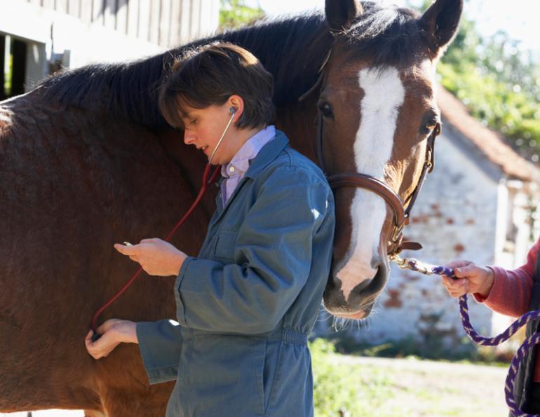
Main article photo: All mammals have occasional extra heartbeats and in the absence of other signs indicating that something is wrong, the extra heartbeats are of no concern. Photographer: MonkeyBusinessImages/iStockphoto.com

Blogs & Podcasts

Advertisement
Related Articles


Advertisement

Featured Stories


Advertisement