Colonic Ulcers in Horses

Updated:
July 3, 2013By Jess Hallas-Kilcoyne
Recent years have seen a dramatic increase in awareness among horse owners of the detrimental effects associated with Equine Gastric Ulcer Syndrome (EGUS). Despite this additional attention paid to the equine digestive system, it remains focused primarily on the horse’s foregut, often ignoring disorders of the hindgut, such as colonic ulcers. The significance of colonic ulcers should not be underestimated, as the underlying causes can potentially lead to very serious health conditions, even death. These cases are rare but even in the absence of such developments colonic ulcers are an affliction that can negatively affect a horse’s quality of life.
Prevalence of Colonic Ulcers
While colonic ulcers seem to occur with less frequency than EGUS, they are still far more prevalent than the average horse owner might think. The data from multiple studies indicates that the incidence of colonic ulcers in horses ranges from 44 percent to a whopping 87 percent, depending on the type and use of the horses.
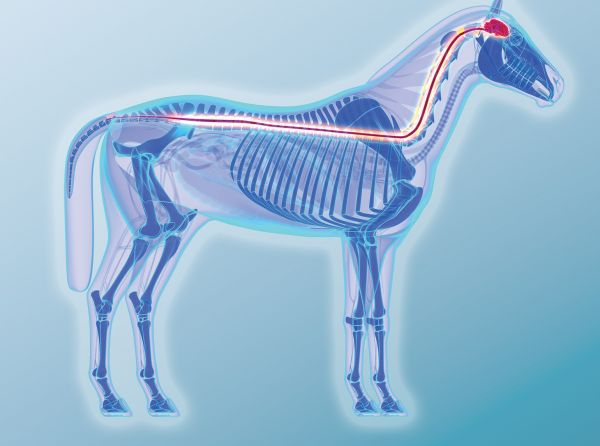
Foregut vs. Hindgut: Anatomy and Function
The Foregut
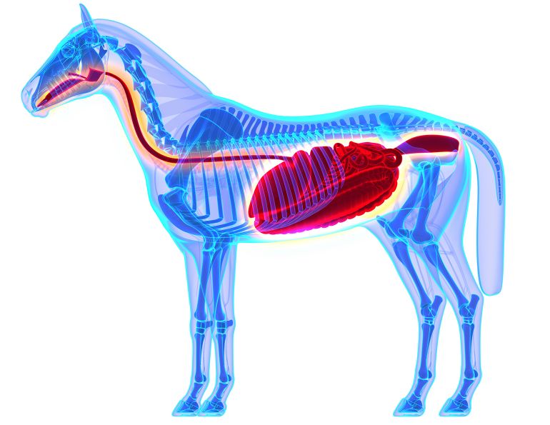
The foregut of the horse is comprised of the esophagus, stomach, and small intestine, and is fairly similar in form and function to that of a human. The esophagus conveys food from the mouth to the stomach, where hydrochloric acid and pepsin (a digestive enzyme) secreted from the stomach walls work to break the food down into smaller particles.
From the stomach, the partially digested food passes into the small intestine. Here, enzymes secreted by the pancreas and by the small intestine itself, as well as bile and lipases from the liver, work to further digest the food material. Once the food has been sufficiently broken down, many nutrients (carbohydrates, amino acids, vitamins, and minerals) are absorbed through the wall of the small intestine. The undigested food material then passes into the large intestine.
The Hindgut
The hindgut consists of the horse’s large intestine, of which there are five portions: the caecum, large colon, small colon, rectum, and anus. The caecum is approximately four feet long, one foot in diameter, and has a capacity of eight to ten gallons. Here, bacterial fermentation coverts the carbohydrates in the undigested fibre material into volatile fatty acids (VFAs) which are then absorbed. This process provides the horse with approximately 70 percent of its energy requirements, and is the reason the horse is called a “hindgut fermenter.”
The large colon is about 12 feet long and 10 inches in diameter, and consists of the right and left ventral and dorsal colons. Microbial digestion continues in the large colon, followed by further absorption of vitamins and minerals.
By the time it reaches the small colon, the food is almost completely digested, and the remaining material is not able to be digested and absorbed by the horse’s body. The primary function of the small colon, which measures about ten feet long and four inches in diameter, is the absorption of excess moisture. This process results in the formation of fecal balls which pass through the rectum and are expelled through the anus.
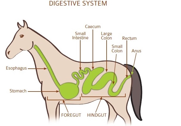
The equine gastrointestinal tract, showing the foregut and the hindgut.
Causes of Colonic Ulcers
Formerly, parasites in the hindgut were thought to be one of the major causes of hindgut issues, including colonic ulcers, but current research indicates that this is very rare.
These days, colonic ulcers in horses are primarily attributed to an ulcerative inflammatory bowel disorder called right dorsal colitis (RDC). Most frequently, RDC is associated with the use of non-steroidal anti-inflammatory drugs (NSAID’s) such as phenylbutazone (Bute). It is not fully understood why the ulcers tend to be localized to the right dorsal colon, but one theory is that the narrowing of the digestive tract after the right dorsal colon results in slower movement of the food (and Bute) and, therefore, prolonged contact between the drug and the right dorsal colon. Another possible cause of RDC is stress, which is thought to trigger the body’s release of corticosteroids that inhibit the function of colon-healthy prostaglandins.
Another potential cause of colonic ulcers is hindgut acidosis (HGA). Hindgut acidosis occurs when large amounts of undigested simple carbohydrates reach the hindgut and are fermented into lactic acid, which causes the pH in the hindgut to fall. The lower pH has a detrimental effect on the colonic mucosa lining and interferes with the hindgut fermentation, reducing the production of VFAs and opening the door for pathogenic or “bad” bacteria to replace the “good” bacteria. It is believed that the effects of the pathogenic bacteria on the weakened mucosa lining may lead to colonic ulceration.
Diagnosis & Clinical Signs
Unfortunately, confirmation of a diagnosis of colonic ulcers cannot be made as easily as that of gastric ulcers, as an equine colonoscopy is an extremely invasive procedure that carries potentially life-threatening risks. If the horse suffers from RDC, ultrasonography may show a thickening of the right dorsal colon, but if the horse’s ulcers are in another region of the colon, there will be no indication on the ultrasound. In many cases, diagnosis of colonic ulcers is based primarily on case history, clinical signs, and response to treatment.
Diagnosis of colonic ulcers based on clinical signs is complicated, made so not only by the fact that many of the symptoms of colonic ulcers are also potential indications of gastric ulcers (although a gastroscopy can be performed to rule out EGUS), but by the possibility that a horse may suffer from gastric and colonic ulcers simultaneously. Clinical signs shared by colonic ulcers and EGUS include colic (mild, intermittent, or acute), poor appetite, weight loss, irritability or poor attitude, low energy, and lackluster performance.
Diarrhea (intermittent or regular) is a symptom that is more typical of an issue in the hindgut, and sensitivity in the girth and flank areas, a “tucked up” abdomen, and reluctance to bend, extend, or collect are all signs that may indicate colonic ulceration.
Treatment & Management
Invariably, the best treatment for colonic ulcers is the adoption of management practices that promote a healthy hindgut. In all cases where colonic ulcers are suspected, discontinuing the use of NSAIDs immediately is strongly recommended. Reducing the duration and intensity of exercise and keeping other stressful activities, such as trailering, to a minimum can also benefit a horse with hindgut issues.
Feeding frequent, low-volume meals throughout the day and increasing dietary fibre will ensure a steady flow of feed matter moving through the horse’s intestinal tract, promoting normal colonic function. Keeping the meals small reduces the mechanical stress placed on the colon. Grain and processed feeds in the horse’s diet should be kept to a minimum to ensure that carbohydrates are digested in the foregut and do not reach the hindgut.
A bulking agent with laxative properties, such as psyllium mucilloid and psyllium hydrophilic mucilloid, can be added to the horse’s diet to ease the passage of material through the hindgut, and may reduce inflammation of the colon.
Feed supplements rich in omega-3 fatty acids, like flax seed, corn, and safflower oils, may also decrease inflammation.
Although no conclusive research has been done into their efficacy, more and more horse owners are now turning to feeding prebiotics and probiotics in an effort to restore and maintain hindgut health. Prebiotics are a non-digestible, dietary fibre that promotes the growth of beneficial bacteria. Probiotics are live microorganisms of the bacteria itself. When ingested by the horse, probiotics add healthy bacteria to the hindgut, and prebiotics nourish the beneficial bacteria and create an environment in which it can flourish.
In terms of medications, while it has yet to be proven that sucralfate is of benefit to the treatment of equine gastric ulcers, it has been used to treat RDC with some success. The prevailing theory is that the sucralfate adheres to the ulcerated tissue in the horse’s colon, forming a barrier over it that protects it from further erosion while stimulating the production of prostaglandins that reduce inflammation and promote healing. Antacids have been shown to be of little benefit to the treatment of colonic ulcers.
Always consult with your veterinarian before making any changes to your horse’s diet and management.
Sources:
1. Overview of Gastric and Colonic Ulcers by Frank M. Andrews, The University of Tennessee, Knoxville, TN; from Advances in Equine Nutrition Vol IV, Kentucky Equine Research, 2009.
2. Colic, Colonic Ulcers and the Equine Digestive Tract, a paper presented by Freedom Health, LLC; last revised August 2011.
3. Right Dorsal Colitis in the Horse by Noreen Galvin, Hugh Dillon, and Frank McGovern, Irish Veterinary Journal, 2004, 57(8), 467-473.
This article is for information purposes only and should not be relied upon as a substitute for professional veterinary advice. No liability will accrue to the publisher or author of the article in the event that a user suffers loss as a result of reliance upon the information.
Main article photo: BLW/Wikimedia Commons
This article originally appeared in the September 2012 issue of Canadian Horse Journal.
Blogs & Podcasts

Advertisement

Related Articles

 and Cushing’s disorder (PPID) phenylbutazone (Bute) horse is rocked back onto its haunches therapeutic hoof boots with pads vitamin e laminitis")

Advertisement

Featured Stories


Advertisement